Skip to content
Patient gallery

Before & after, consented and unretouched.

Every case here is published with the patient's written consent and shown exactly as photographed — nothing is retouched, smoothed or restaged. This is reconstructive work, so the “before” is often an injury or a tumour rather than a cosmetic concern.

Some photographs on this page are graphic. Surgical wounds, an amputated limb and an operative specimen appear in these cases. Those images stay hidden behind a tap, and are not loaded at all until you ask to see them. Identifying features are obscured unless a patient has signed explicit identification consent.

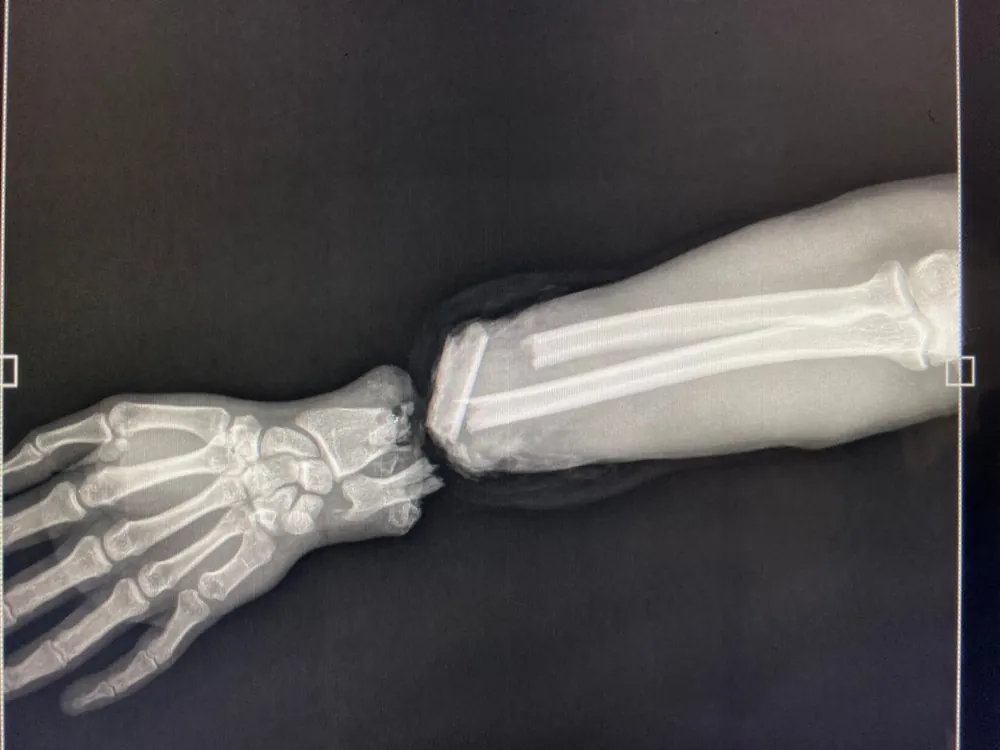
Radiograph on arrival — the hand is completely separated at the wrist.
Before
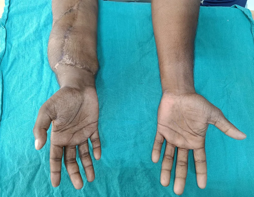
Nine months after surgery, alongside the uninjured hand.
After · 9 months

This case includes graphic clinical photographs. They stay hidden until you choose to view them.

Hand & Limb

Hand replantation at the wrist

Presentation
A complete traumatic amputation of the hand through the wrist. The hand arrived separated from the forearm; the radiograph shows the full discontinuity that had to be bridged.
Approach
Emergency replantation. The skeleton was fixed first to give the repair a stable frame, then the arteries and veins were reconnected under the microscope to restore circulation, and the nerves and tendons repaired to give the hand a route back to movement and sensation.
Outcome
The hand survived. At nine months the wrist had healed, the fingers extended and flexed actively, and the replanted hand sat level with the uninjured one.
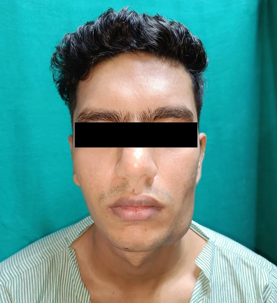
Before surgery — fullness of the lower jaw distorting facial symmetry.
Before
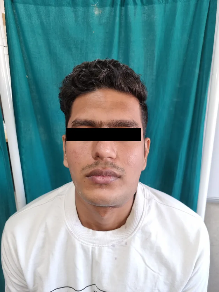
Nine weeks after surgery — jawline and facial symmetry restored.
After · 9 weeks

This case includes graphic clinical photographs. They stay hidden until you choose to view them.

Craniofacial

Jaw tumour resection and reconstruction

Presentation
A tumour of the lower jaw, presenting with a visible swelling that had pushed the face out of symmetry and left the patient unable to open the mouth fully.
Approach
The tumour-bearing segment of the jaw was resected and the jaw reconstructed in the same operation, so the patient did not spend the healing period without a mandible.
Outcome
Facial symmetry was restored and the patient regained a full range of mouth opening — visible in the post-operative photographs taken nine weeks after surgery.
Before
After · 5 weeks

This case includes graphic clinical photographs. They stay hidden until you choose to view them.

Hand & Limb

Heel reconstruction with a flap

Presentation
A large, long-standing wound of the heel with dead tissue at its base, sited on the part of the foot that has to take the body's weight. Wounds in this position do not close on their own, and left alone they put the limb at risk.
Approach
The dead tissue was removed down to a clean base and the defect resurfaced with a flap — living tissue with its own blood supply, moved in to give the heel padding durable enough to walk on.
Outcome
The flap took and the heel healed with a stable, weight-bearing surface. The limb was preserved.
Results vary from patient to patient. The cases shown here were selected to illustrate what reconstruction can achieve, not to promise a particular outcome — what is achievable in your case depends on your anatomy, the injury or diagnosis, and how early treatment begins. Dr. Dave will talk you through a realistic expectation at consultation.
Begin your consultation

A private conversation about what's possible.

Forty-five minutes with Dr. Dave. A clinical examination. 3D imaging where relevant. A written plan and transparent quote. No obligation, no upsell — just an honest discussion of your options.

Book an appointment