

Craniofacial
Jaw tumour resection and reconstruction
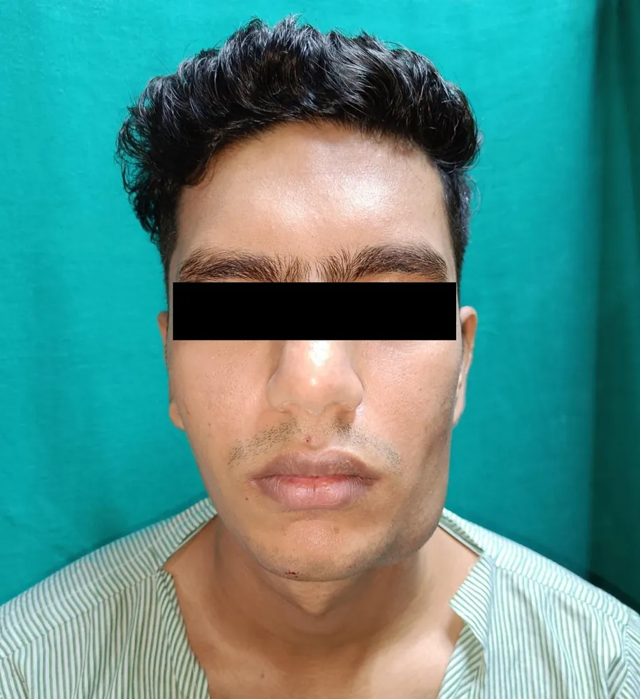
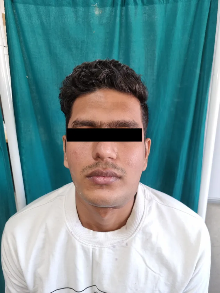
Facial symmetry was restored and the patient regained a full range of mouth opening — visible in the post-operative photographs taken nine weeks after surgery.
9 weeks post-op
Treatment of the bones of the face and jaws — facial fractures, corrective jaw (orthognathic) surgery, TMJ problems and facial deformity — to restore both function and facial harmony.

Maxillofacial surgery is the surgery of the facial skeleton — the bones of the nose, cheek, eye socket and the upper and lower jaws — together with the soft tissues and joints that move them. It sits at the crossroads of trauma, function and appearance, because the face is at once a structure that must work and a structure that must look like itself. The work falls into a few broad strands. Facial trauma is the rebuilding of bones broken in road accidents, falls and assaults — fractures of the nasal bones, the cheekbone (zygoma), the floor and walls of the eye socket (orbit), the lower jaw (mandible) and the midface (the Le Fort pattern of fractures) — where the goal is to set the bones back to their original position and hold them with fine titanium plates and screws. Corrective jaw surgery, or orthognathic surgery, repositions jaws that are too far forward, too far back or asymmetric, correcting the bite (malocclusion) and the facial imbalance that comes with it — planned in close coordination with an orthodontist. Alongside these sit the management of TMJ (jaw joint) disorders and the correction of established facial deformity. Throughout, Dr. Aniket Dave plans to a single principle: function first — a bite that meets, an airway that breathes, eyes that align and track — with facial harmony restored in the same breath. Modern fixation and 3D planning make outcomes more predictable, and trauma readiness means a broken face can be addressed promptly in Ahmedabad rather than left to set wrongly.
Maxillofacial surgery is considered when the bones, joints or bite of the face need to be repaired or repositioned.
A broken or misaligned facial skeleton is corrected to anatomy, not to approximation. For trauma, Dr. Dave reduces fractures back to their pre-injury position and fixes them with low-profile titanium plates and screws placed through hidden incisions wherever possible — inside the mouth, in the eyelid crease, in the hairline — so the repair is functional and discreet. For corrective jaw surgery, planning is the operation: dental models, a CT scan and 3D virtual surgical planning are used to rehearse the move, design cutting guides and predict the bite and the profile before theatre, with the orthodontist aligning the teeth before and after. The aim is always twofold — restore what the face must do (bite, breathe, see, speak) and restore how it should sit (symmetry, proportion, the patient's own features). Surgery is carried out under consultant anaesthesia, and the trauma pathway is built to address fractures promptly before bone begins to set in the wrong place.
How a maxillofacial case proceeds, in broad terms.
Clinical examination, dental occlusion review and CT imaging to map the fractures or plan the jaw movement; photographs from standard angles.
For jaw surgery, 3D virtual planning, dental models and orthodontic alignment; for trauma, a fixation plan and timing decision.
Administered by a consultant anaesthetist; the airway is secured to allow work in and around the mouth. You sleep throughout.
Exposure via intra-oral, eyelid-crease or hairline incisions wherever possible to avoid visible facial scars.
Fractured bones are reduced to their correct position, or the jaws are cut and moved (osteotomy) to the planned occlusion.
The bones are held with low-profile titanium plates and screws; the bite is checked and secured before closure.
Layered closure, dissolvable intra-oral sutures where used, and recovery in a monitored setting before the ward.
Facial swelling peaks; managed with cold compresses, head elevation and analgesia. Soft or liquid diet; oral hygiene as instructed.
Swelling begins to settle. Skin sutures removed if used. Many patients return to light desk work as comfort allows.
Most visible bruising resolves. Soft diet continues. Jaw-surgery patients begin guided jaw exercises or elastics per plan.
Bone fixation is consolidating; diet is gradually advanced. Trauma patients reviewed for healing on imaging if indicated.
Bone heals fully and the final bite and facial contour settle. Orthodontic finishing continues after jaw surgery; residual swelling fades.
Published with the patient's written consent and shown unretouched. Graphic photographs stay hidden until you choose to view them.
Facial symmetry was restored and the patient regained a full range of mouth opening — visible in the post-operative photographs taken nine weeks after surgery.
Trauma cases are often insured; orthognathic surgery varies with complexity.
Maxillofacial pricing in Ahmedabad ranges widely because the work itself ranges widely — a single isolated jaw fracture is a different undertaking from a two-jaw orthognathic correction with 3D planning, or a panfacial reconstruction after major trauma. Theatre time, the number of fractures or osteotomies, the volume of titanium hardware, imaging and planning, and hospital stay all move the figure. Importantly, facial trauma is commonly covered by health insurance, and we help patients and families with pre-authorisation and documentation. Orthognathic surgery is quoted transparently after planning, when the scope is clear.
Maxillofacial surgery is well-established and safe in trained hands, but it carries real risks that Dr. Dave discusses frankly before surgery.
Forty-five minutes with Dr. Dave. A clinical examination. 3D imaging where relevant. A written plan and transparent quote. No obligation, no upsell — just an honest discussion of your options.


