

Hand & Limb
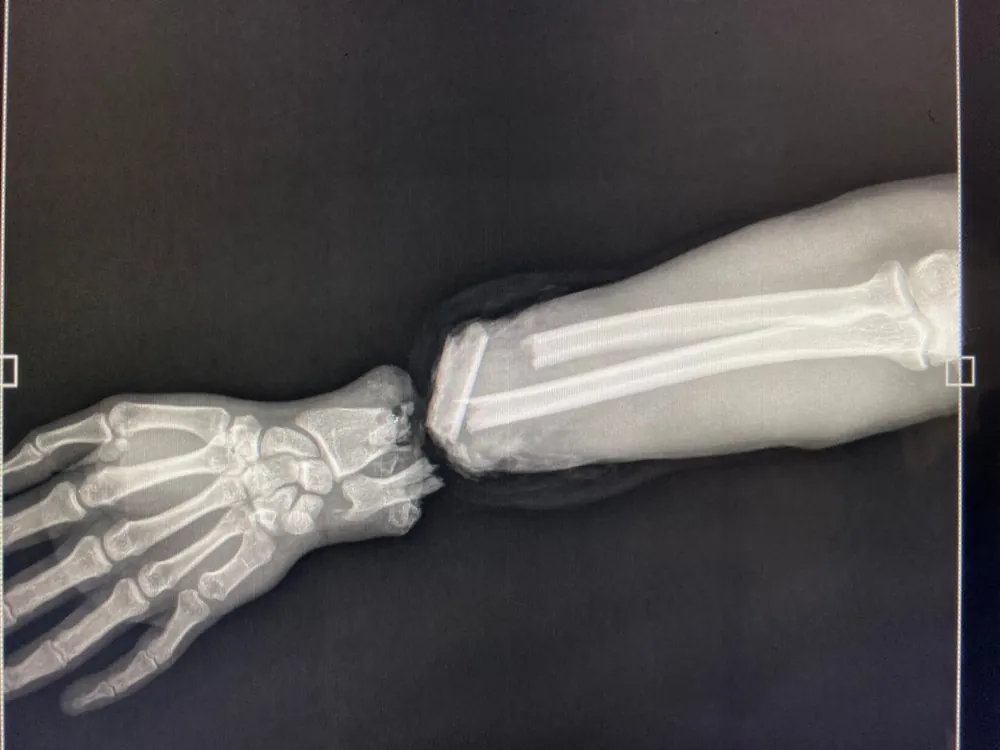
Hand replantation at the wrist
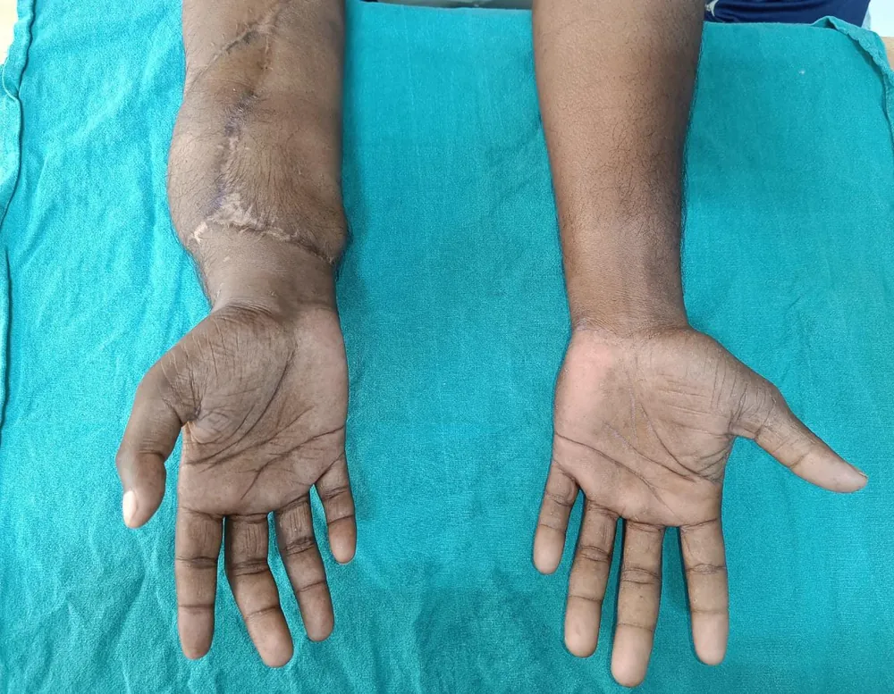
The hand survived. At nine months the wrist had healed, the fingers extended and flexed actively, and the replanted hand sat level with the uninjured one.
9 months post-op
Free flap reconstruction for cancer, major trauma and chronic wounds — vessels under 2 mm reconnected under the operating microscope to restore both form and function.

Reconstructive microsurgery is the most technically demanding discipline in plastic surgery — the rebuilding of a defect by moving living tissue from one part of the body to another and reconnecting its blood supply under the operating microscope. This is free tissue transfer, or a free flap: skin, fat, muscle or bone is detached completely from its original site, transplanted to the defect, and its artery and vein — often less than 2 mm across — are sutured to recipient vessels using thread finer than a human hair. Within minutes the tissue is alive again in its new home. It is the technique that makes the impossible routine: rebuilding a jaw after oral cancer with a section of fibula, reconstructing a breast with the patient's own abdominal tissue, covering an exposed tibia after a road accident, or filling a chronic wound eroded by osteomyelitis. Dr. Aniket Dave's core specialty is microsurgical reconstruction, and he works hand-in-hand with oncologists, trauma teams and orthopaedic surgeons across Ahmedabad. The guiding principle is never closure alone — it is the restoration of both form and function, so that a reconstructed jaw can chew, a reconstructed limb can bear weight, and a reconstructed face can be lived in without apology. Free flaps demand a surgeon comfortable at the highest acuity and a system built to monitor a flap's blood supply hour by hour in the days after surgery.
Reconstructive microsurgery is considered when a defect is too large or too complex for local tissue alone.
A free flap is rarely a solo undertaking. For head & neck cancer, Dr. Dave plans the reconstruction in the same sitting as the resecting surgeon, so that the fibula or radial forearm flap is shaped to the exact defect the moment it is created. For trauma, the timing of flap cover is critical — early definitive cover protects exposed bone and lowers infection risk. The flap is chosen for the job, not for habit: a thin, pliable radial forearm for the inside of the mouth; a bulky ALT or gracilis for a deep cavity; a fibula when bone is needed. Every case is rehearsed against imaging and a clear functional goal. Surgery is performed under consultant anaesthesia with the operating microscope, and the flap is monitored intensively afterwards because the first 72 hours decide its survival.
How a free flap reconstruction unfolds.
Imaging, vessel mapping and a joint plan with the oncology or trauma team to match the right flap to the defect.
Administered by a consultant anaesthetist for a long, stable operation; you sleep throughout.
The tumour bed or wound is cleared, debrided of dead tissue, and recipient artery and vein are identified.
The flap — ALT, DIEP, fibula, gracilis or radial forearm — is raised on its feeding vessels and tailored to the defect.
Under the operating microscope, the flap's vessels (often under 2 mm) are sutured to recipient vessels and blood flow is restored.
The flap is positioned, contoured and secured; bone segments are plated where a jaw or limb is being rebuilt.
The donor site is closed, drains placed, and the flap is monitored hourly for colour, warmth and Doppler signal.
Intensive flap monitoring in a high-dependency setting. Strict positioning, warmth and hydration to protect the new blood supply.
Drains removed as output settles. Mobilisation begins. Donor-site care and early swallowing or weight-bearing assessment by site.
Discharge for most uncomplicated flaps. Wound healing consolidates; dressings simplify. Light daily activity resumes.
Return to most normal activity. Function-focused rehabilitation — speech and swallow therapy, or graded limb loading — continues.
Flap softens and settles. Secondary refinements (debulking, contouring) are planned once tissues are stable.
Published with the patient's written consent and shown unretouched. Graphic photographs stay hidden until you choose to view them.
The hand survived. At nine months the wrist had healed, the fingers extended and flexed actively, and the replanted hand sat level with the uninjured one.
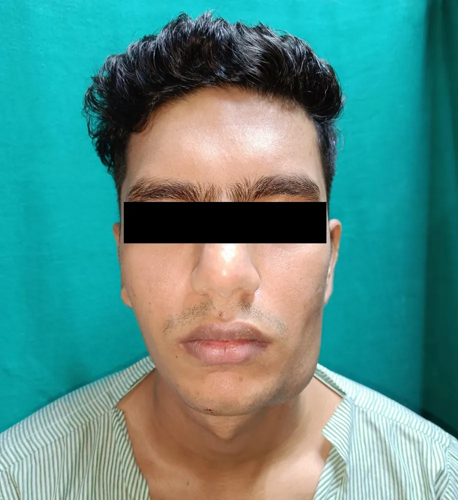
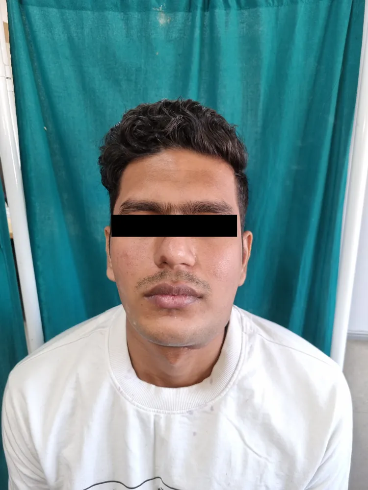
Facial symmetry was restored and the patient regained a full range of mouth opening — visible in the post-operative photographs taken nine weeks after surgery.
The flap took and the heel healed with a stable, weight-bearing surface. The limb was preserved.
Highly variable; oncologic and trauma reconstruction is often insured.
Free flap reconstruction in Ahmedabad spans a wide range because no two reconstructions are alike — a single soft-tissue flap for a wound is a different undertaking from a fibula jaw reconstruction with plating after cancer. Theatre time, microsurgical complexity, hospital and high-dependency stay, and any bone or hardware all move the figure. Importantly, reconstruction following cancer resection or major trauma is commonly covered by health insurance, and we help patients and families navigate pre-authorisation and paperwork.
Microsurgery is reliable in trained hands, but free tissue transfer carries real risks that Dr. Dave discusses frankly before surgery.
Forty-five minutes with Dr. Dave. A clinical examination. 3D imaging where relevant. A written plan and transparent quote. No obligation, no upsell — just an honest discussion of your options.


